Red Infarct
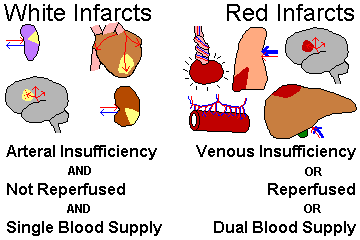
Hemorrhagic. Occurs in loose tissues with collaterals (i.e. liver, lungs, intestine) or following reperfusion (the return of blood flow)
Pale Infarct
Occurs in solid tissue with single blood supply: heart, kidney, and spleen.
Hypovolemic/cardiogenic shock
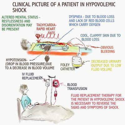
Can be due to a low output failure and high transpulmonary resistance. Or the cardiac output may just be low. The patient will be cold and clammy due to vasoconstriction.
Septic shock
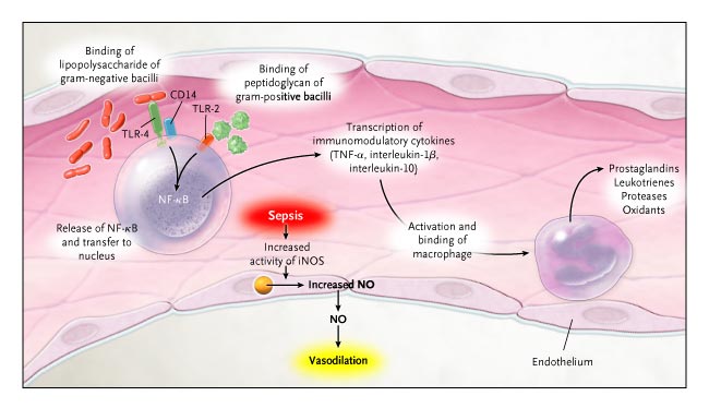
There is a high-output failure coupled with the low transpulmonary pressure. Arterioles are all dilated and there is tremendous venous return. The patient is HOT!
Atrophy
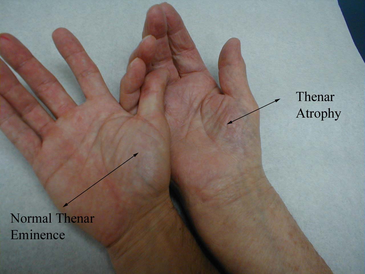
Atrophy is a reduction in the size and/or number of cells. It can occur from low hormone levels, loss of innervation, ischemia, not enough nutrients, or a local increase in pressure (nephrolithiasis). Also, an occlusion of a secretory duct as in cystic fibrosis can atrophy areas.
Inflammation
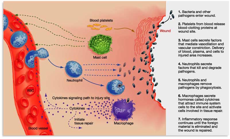
Rubor, dolor, calor, tumor (swelling), functio laesa (loss of function). The vasculature becomes highly permeable, dilates, and there is some endothelial injury. Neutrophils extravasate and phagocytose, degranulate, and release inflammatory mediators. Depending on whether it is acute or chronic, different cells will predominate.
Acute: you have neutrophils, eosinophils, and antibodies. It comes on in seconds and may last a few minutes or even a few days. It may go away completely or leave an abscess, or turn into chronic inflammation.
Chronic: Mostly mononuclear cells with lots of destruction and repair. Blood vessels proliferate and fibrosis ensues. Granulomas form which are just nodular collections of epithelioid macrophages and giant cells. There will be scarring and amyloidosis (deposit of protein nodule)
Free Radical Injury
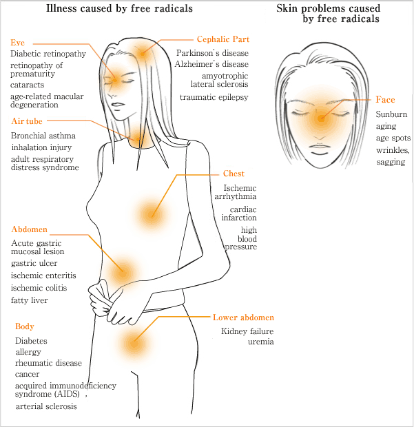
Free radicals cause damage in three ways: lipid peroxidation, protein modification, and DNA breakage. This can happen by various methods including exposure to radiation, the phase I metabolism of drugs, redox reactions, nitric oxide, transition metals, and the leukocytes oxidative burst.
The battle against free rads is fought by several enzymes including catalase, superoxide dismutase, glutathione peroxidase, and also by spontaneous decay and several vitamins that act as antioxidants like A, C, and E, or ACE!
Free radicals are associated with a lot of pathology: Retinopathy of prematurity, Bronchopulmonary dysplasia, Carbon tetrachloride which leads to fatty liver and necrosis, Acetaminophen overdoses which causes fulminant hepatitis, iron overload (hemochromatosis), and Reperfusion after anoxia usually after thrombolytic therapy i.e. superoxide.
Granulomatous diseases
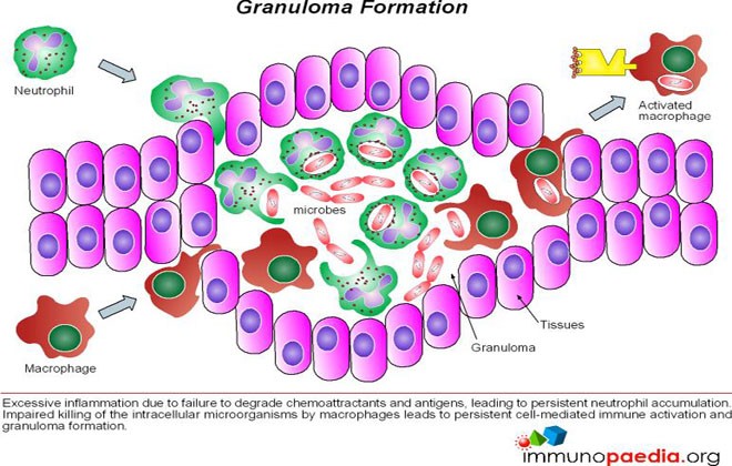
In Granulomatous diseases, Th1 cells secrete gamma-interferon to activate macrophages. Then, the macrophages release TNF-alpha which is responsible for the granuloma formation. Careful when giving TNF-alpha suppressors because they can lead to breakdown and dissemination of formed granulomas.
Diseases that cause granulomas to form are: Mycobacterium tuberculosis, fungal infections, Treponema pallidum which causes syphilis, M. leprae or leprosy, Bartonella henselae which is cat scratch disease, sarcoidosis, Crohns, Wegeners, Churg-Strauss syndrome, and Berylliosis
Diseases that cause granulomas to form are: Mycobacterium tuberculosis, fungal infections, Treponema pallidum which causes syphilis, M. leprae or leprosy, Bartonella henselae which is cat scratch disease, sarcoidosis, Crohns, Wegeners, Churg-Strauss syndrome, and Berylliosis
Iron poisoning
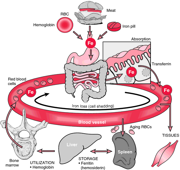
One of the leading causes of fatal toxicity in children.
Membrane lipids of cells undergo peroxidation.
Acutely you will find the patient suffering from gastric bleeding. If the condition is chronic, there will be metabolic acidosis, and scarring in GI leading to obstruction.
Membrane lipids of cells undergo peroxidation.
Acutely you will find the patient suffering from gastric bleeding. If the condition is chronic, there will be metabolic acidosis, and scarring in GI leading to obstruction.
Amyloidosis
This is an abnormal aggregation of proteins or their fragments into beta pleated sheets. The cells become damaged from this and apoptose. The tissue will look waxy.
There are many types of amyloidosis:
AL or primary is due to the Ig light chains and can occur in the plasma be associated with multiple myeloma (cancer in the bone marrow). Several organs are effected.
AA 0r secondary is when the protein is serum Amyloid A and is seen with RA, IBD, spondyloarthropathy, and chronic infections.
This also happens to patients undergoing dialysis and shows up as carpal tunnel.
It can be heritable due to a gene mutation such as in pre albumin.
It can come with age (senile) due to TTR deposition in the myocardium.
And finally, it can be organ specific as often seen with Alzheimers patients from amyloid-beta cleaved from the amyloid precursor protein.
Neoplastic progression

The hallmarks of cancer include: evading apoptosis, self sufficient growth signaling, insensitive to anti-growth signals, sustained angiogenesis, limitless replicative potential, metastasis, and tissue invasion.
What happens is first, there is hyperplasia, where the cells increase in number and also a dysplasia because these new cells no longer resemble the original.
Then, this is when you have a carcinoma in situ. The cells have yet in invade the basement membrane, they have huge nucleuses and not too much cytoplasm.
Using collagenases and hydrolases, the cells can now invade the basement tissue and have the potential to metastasize if they reach a vessel.
Once metastatic, all they have to do is avoid immune attack.
Anaplasia
These are irreversible undifferentiated malignant neoplasms. They resemble the primitive cells of the same tissue.
Neoplasia
This is an uncontrolled proliferation that may either be benign or malignant.
Desmoplasia
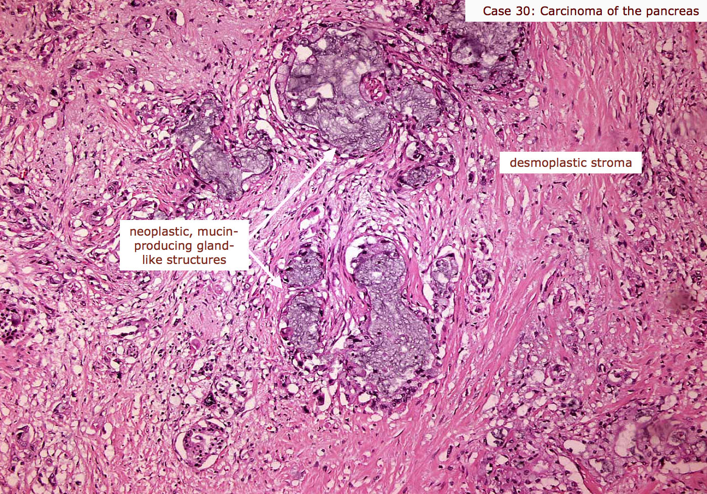
This is the fibrous tissue that forms with a neoplasm.
Epithelial tumor
If benign: Adenoma or papilloma
Malignant: Adenocarcinoma or papillary carcinoma
Malignant: Adenocarcinoma or papillary carcinoma
Blood cell tumor
Only malignancy exists and is termed Leukemia or lymphoma.
Blood vessel tumor
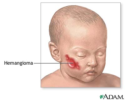
Benign: Hemangioma
Malignant: Angiosarcoma
Malignant: Angiosarcoma
Smooth muscle tumor
Benign: Leiomyoma
Malignant: Leiomyosarcoma
Malignant: Leiomyosarcoma
Striated muscle tumor
Benign: Rhabdomyoma
Malignant: Rhabdomyosarcoma
Malignant: Rhabdomyosarcoma
Connective tissue tumor
Benign: Fibroma
Malignant: Fibrosarcoma
Malignant: Fibrosarcoma
Bone tumor
Benign: Osteoma
Malignant: Osteosarcoma
Malignant: Osteosarcoma
Fat tumor
Benign: Lipoma
Malignant: Liposarcoma
Malignant: Liposarcoma
Cachexia
This term describes the weight loss, muscle atrophy, and fatigue often seen with chronic diseases such as AIDS and cancer. It is mediated by TNF-alpha a.k.a. "cachectin", IFN-gamma, and IL-6.
The following are diseases and their associated neoplasms (16)
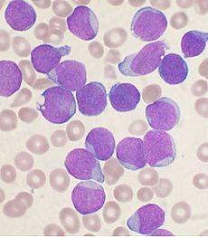
Down syndrome
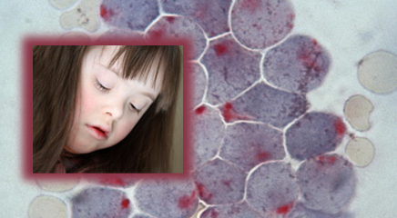
ALL and AML (acute myeloid leukemia)
Xeroderma pigmentosum, albinism
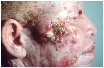
Melanoma, basal cell carcinoma, and squamous cell carcinomas of the skin.
Chronic atrophic gastritis, pernicious anemia, postsurgical gastric remnants
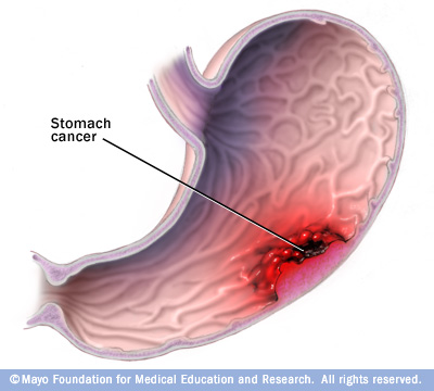
Gastric adenocarcinoma
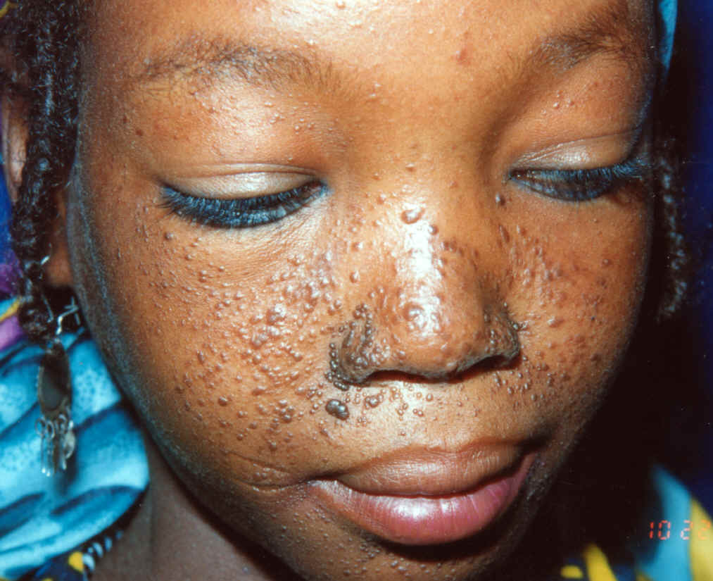
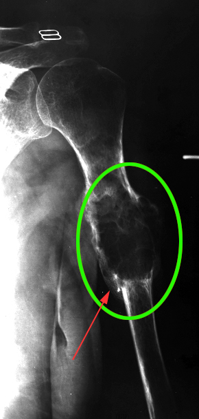
Tuberous sclerosis (facial angiofibroma, seizures, mental retardation)
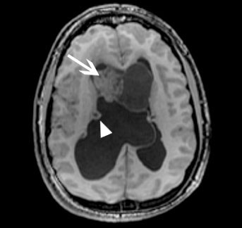
Giant cell astrocytoma (pictured), renal angiomyolipoma, and cardiac rhabdomyoma
